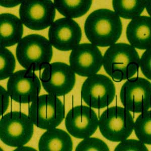
Fluorescent microspheres refer to particles with diameters ranging from nanometers to micrometers (0 Solid particles loaded with fluorescent substances within the range of 01-10 μ m, which can be excited to emit fluorescence by external energy stimulation. Lateral flow assay (LFA) has developed rapidly in recent years, with advantages such as simplicity, speed, independence from large instruments and equipment, and low cost. It is widely used in real-time inspection, on-site inspection, and home self inspection.

Fluorescent microspheres, as a special type of functional microsphere, not only possess the properties of inorganic and organic substances, but also emit fluorescence under external energy stimulation. The recently developed fluorescent microspheres have uniform particle size, good monodispersity, good stability, and high luminescence efficiency. The surface curvature of the microspheres is conducive to the optimal reaction state of the exposed surface of the antigen determinant cluster and antibody binding site.
Therefore, they have a wide range of applications in many fields, such as biochemistry, biomedicine, clinical medicine, gene analysis, and optical instruments. Among them, the application of fluorescent microspheres in medicine and biology is particularly important. The loading of fluorescent microspheres on microspheres can be divided into four methods: swelling adsorption, embedding, chemical bonding, and copolymerization
01 Application in marking and tracing
Fluorescent microspheres prepared by introducing one or more fluorescent substances on the surface or inside of polymer microspheres were initially used as fluorescent standard microspheres for calibrating flow cytometers and fluorescence microscopes, and gradually applied to cell labeling, biomolecule labeling, and tracking under active conditions. They can also immobilize protein molecules and track their functionalization process.
02 Application in detection
Mixing fluorescent microspheres with ligands that can bind to the analyte on the surface with radiolabeled analyte, the analyte that can bind to the microspheres in the system can be excited by their radiation to produce fluorescence, while the analyte that cannot bind to the microspheres can be submerged in water due to their distance and cannot be excited by their radiation, allowing for the detection of non separated products.
Moreover, fluorescent microspheres can carry many fluorescent molecules, and weaker stimuli can trigger stronger signals, so only a small amount of low-energy radiation is needed to generate fluorescent signals, avoiding the radiation hazards caused by the use of traditional radioactive microspheres and reducing costs without losing detection sensitivity.
03 Applications in Immunoassay and Drug Screening
At present, the most widely used and promising application of fluorescent microspheres is in the biomedical field, and the industrialized fluorescent microspheres abroad are mainly used in this field. It mainly includes high-throughput immunoassay, drug screening, immobilized enzymes, etc. Fluorescent microspheres can achieve sensitive, rapid, and efficient detection of various microorganisms. Carboxylated fluorescent microspheres can be covalently coupled with monoclonal antibodies, and corresponding fluorescent microsphere immunochromatographic test strips can be prepared through a double antibody sandwich reaction mode for detection.
Fluorescent microspheres serve as immobilized carriers for different detection targets in biological sample detection. They bind to antibodies or antigens through physical adsorption or covalent binding, and can detect corresponding antigens or antibodies in body fluids through agglutination testing. This method has been widely used in the early stages due to its simplicity and sensitivity, and different fluorescent microspheres correspond to different capture antibodies, which can recognize different antigens and achieve qualitative and quantitative detection of multiple test antigens.
04 Application in immobilized enzyme and gene research
Enzymes can be fixed on fluorescent microspheres through physical adsorption or chemical bonding. Due to the fixed three-dimensional morphology of enzymes, they not only have high pH stability, thermal stability, and storage stability, but also are easy to separate from reactants, can be reused, and improve efficiency.
Therefore, immobilizing enzymes or other bioactive substances on microsphere carriers is more conducive to their functional performance. Meanwhile, fluorescent microspheres can also be connected to targets such as genes through physical adsorption or chemical bonding. By binding these targets to corresponding markers, detection and analysis can be carried out.
In addition, fluorescent microspheres can also be used as standards for calibrating optical instruments due to their stable morphological structure and efficient luminescence efficiency.
Time resolved fluorescence immunoassay is one of the most advanced immunoassay methods in contemporary times. Combined with the amplification effect of nano signals, it has broad application prospects and is suitable for the development of high-sensitivity immunoassay kits for tumor markers, hormones, and other factors.
Due to the low luminescence efficiency of rare earth ions themselves, when rare earth ions form complexes with ligands with high absorption coefficients, the ligands absorb laser and transition to an excited state, transferring energy to the rare earth ions. When the rare earth ions receive the transferred energy, they are excited to the resonance energy level and emit fluorescence during the transition back to the ground state, exhibiting strong rare earth ion characteristic fluorescence.
Based on the above microsphere structure and luminescence mechanism, time-resolved fluorescent microspheres have the following characteristics compared to ordinary fluorescent microspheres:
(1) Internal embedding process – dye free surface for easy coupling
(2) Fluorescence intensity – higher than ordinary red or green fluorescent microspheres
(3) Stokes displacement – larger than ordinary fluorescent microspheres (mostly above 250nm)
(4) Half life – long fluorescence lifetime, strong anti-interference ability
The time-resolved fluorescence lifetime is usually over 100 μ s, which is several orders of magnitude higher than the fluorescence lifetime of ordinary fluorescent microspheres. This is because the luminescence of rare earth complexes is caused by the energy transfer of the excited state of the ligand
(5) Stability – Rare earth ion complexes have higher stability than ordinary fluorescence and more stable inter batch differences
At present, the most common time-resolved fluorescent microspheres is embedded with rare earth element europium (Eu), which is mainly used in the development of fluorescent quantitative immunochromatographic test strips for POCT rapid detection, food safety testing (including pesticide residues, drugs, etc.), etc. Combined with a fluorescence quantitative detector, rapid quantitative detection of different indicators can be achieved.
