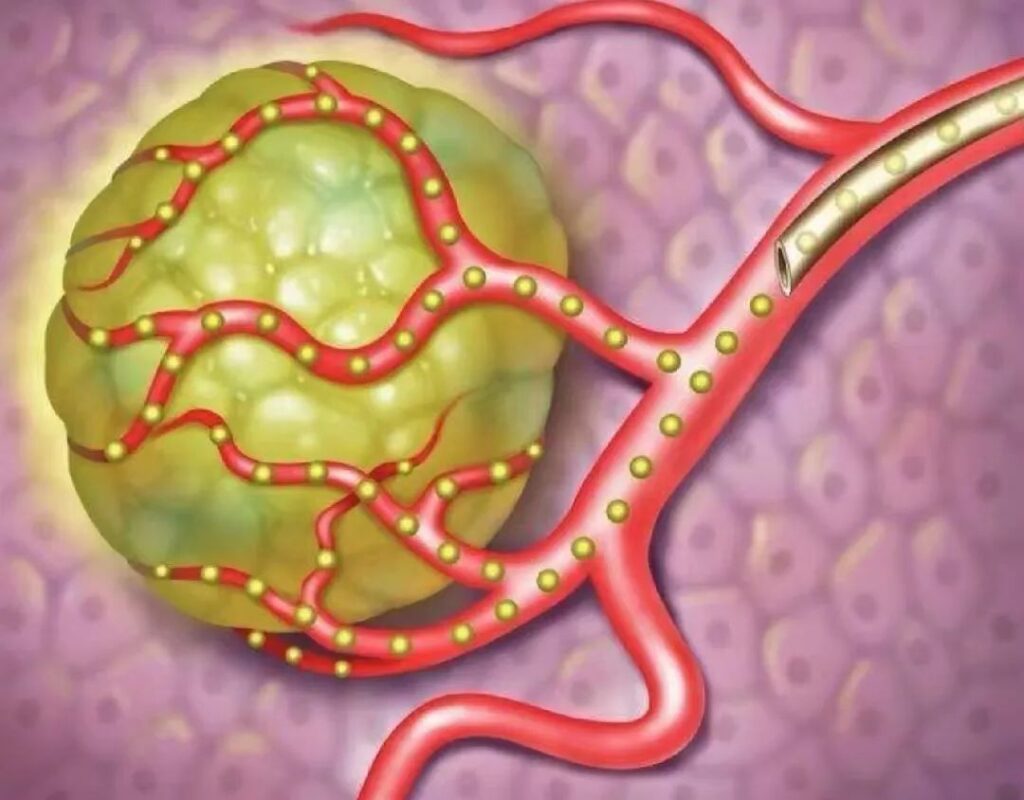
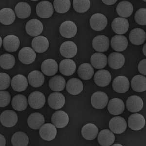
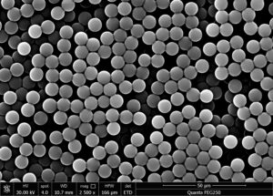
Microspheres for drug delivery refer to spherical particles formed by the dissolution or dispersion of drugs in polymer matrix materials, with a particle size generally between 1-250 μm. They can be used for different administration routes such as intravenous injection, oral administration, local administration through the cavity, or subcutaneous implantation.
Microsphere formulations can slowly release drugs, thereby reducing the frequency of administration, lowering fluctuations in blood drug concentration, and exerting long-term therapeutic effects. Introducing magnetic substances into microspheres or modifying carrier materials can enable microspheres to magnetically or actively target lesions, increase effective blood drug concentration at the target site, and reduce systemic toxic side effects of drugs. Introducing microspheres into tumor arteries can kill tumor cells through embolization therapy, blocking the tumor’s nutrition and blood supply while releasing drugs, further increasing the therapeutic effect. In addition, microsphere formulations can also mask the unpleasant odor of drugs, reduce irritation, and improve drug stability.
In particular, microspheres drug delivery systems can utilize their own advantages to achieve local delivery of large molecule drugs such as small molecule drugs, peptides, proteins, and engineered cells.
For example, microspheres can promote the adhesion and proliferation of bone marrow mesenchymal stem cells (BMSCs), and implantation of microspheres into bone defects can promote bone formation; When used for transarter arterial chemoembolization (TACE), microspheres can be prepared as needed according to the vessel diameter and treatment requirements to achieve more precise embolization effects; Prepare microspheres into porous and multi-layered structures, loading different contents while forming a long-lasting drug reservoir in vivo; Utilize the differences in degradation rates of different materials to achieve sequential or gradient drug release.
The application of microsphere preparations in the medical field has made significant progress, with multiple products successfully launched, providing innovative treatment strategies for various diseases.
1. Preparation method of microspheres drug delivery
The preparation process of microspheres for drug delivery usually includes four steps: dispersion, solidification, washing, and drying. Dispersion refers to the uniform distribution of drugs into a polymer matrix through emulsification, control of solute solubility, and other methods to form microsphere structures. Curing involves fixing the morphology and structure of microspheres through physical methods (such as solvent evaporation, temperature changes) or chemical methods (such as cross-linking reactions), and then washing and drying to remove impurities, resulting in microsphere particles that can be stored. This article classifies drug loaded microspheres based on different preparation methods, analyzes the core processes of each method.
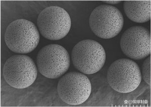
2.Clinical Application of Microspheres Drug Delivery System
2.1 Local treatment for malignant tumors
Malignant tumors (cancer) are a major disease that threatens social stability and economic development in China. In 2022, there will be approximately 4.8 million new cases of cancer and 2.5 million cancer deaths in China. The anti-tumor drugs currently used in clinical practice mainly include molecular targeted drugs, chemotherapy drugs, immunotherapy drugs, and cell therapy. Encapsulating anti-tumor drugs in microspheres can not only delay the release rate, continuously kill tumor cells, overcome the occurrence of drug resistance and administration frequency, but also reduce adverse reactions through local or in situ administration strategies.
Xi et al. prepared porous polylactic acid (PLA) microspheres by double lotion method and solvent extraction method. By utilizing the self-healing properties of PLA, mild infrared light irradiation causes an increase in temperature, transforming PLA from a glassy state to an rubbery state and triggering spontaneous rearrangement of polymer chains. This porous microsphere will undergo healing, loading antigen molecules into the microsphere to achieve sustained release effect.
Microspheres can also encapsulate various contrast agents for visualization in vivo. Zhang et al. combined electrospinning, homogenization and electric spray technology to prepare hyaluronic acid functionalized drug loaded fiber microspheres. The Gd3+chelated on the microspheres can achieve magnetic resonance imaging of tumors for at least 5 days.
To prevent metastasis and recurrence after solid tumor resection, adjuvant therapy such as chemotherapy or radiotherapy is often used postoperatively. However, the concentration of chemotherapy drugs reaching the target organ after systemic administration is limited, and reaching a certain drug concentration requires a larger dose, which can lead to systemic toxic side effects. Zhong et al. used microfluidic method and electric spray method to prepare calcium alginate microspheres containing multiple methacryloyl gelatin (GelMA) microspheres.

The calcium alginate microspheres are filled in situ at the site of tumor resection. The rapid degradation of calcium alginate microspheres results in the rapid release of doxorubicin to kill residual tumor cells, while GelMA microspheres degrade slowly and continuously release the encapsulated liver regeneration promoter. GelMA microspheres can also serve as a liver cell regeneration scaffold to promote liver regeneration.
In recent years, chimeric antigen receptor T cells (CAR-T) therapy has produced effective and long-lasting clinical responses in the treatment of hematological malignancies, and is expected to change the current status of hematological cancer treatment. But currently, CAR-T therapy has limited efficacy in solid tumors, mainly because the dense extracellular matrix and abnormal vascular system of solid tumors limit the tumor infiltration of CAR-T cells.
Inspired by the physiological process of T cell proliferation in lymph nodes, Liao et al. prepared PLGA microspheres artificial lymph node scaffolds using microfluidic method for loading CAR-T cells and encapsulating multiple cytokines, simulating the key signaling molecules provided by antigen-presenting cells (APCs) to activate T cells.
2.2 Used for treating orthopedic diseases such as bone defect repair
The microenvironment of bone injury is characterized by inflammation, acidity, and high expression of reactive oxygen species (ROS). Drugs such as cytokines play a crucial role in bone defect repair, but their application is limited by the inability of cytokines to maintain long-term activity in complex body environments. Microspheres can provide a stable microenvironment for it, which can retain its activity for a long time and achieve sustained release effect. And the injectability of microspheres allows them to be implanted into patients’ bodies to fill irregular bone defect areas. Song et al. loaded manganese dioxide (MnO2) nanoparticles and bone morphogenetic protein-2 (BMP-2) onto PLGA microspheres, The responsive on-demand release of drugs was achieved using low-frequency ultrasound technology.
Although bone tissue has a certain regenerative ability, for larger bone defects that exceed the self-healing ability of bone tissue, bone graft implantation is usually required to achieve effective treatment results. Hao et al. prepared GelMA/methacryloyl hyaluronic acid (HAMA) microspheres encapsulating decalcified bone matrix (DBM) powder and vascular endothelial growth factor using microfluidic method, and loaded the microspheres into DBM scaffolds for implantation into bone defects.
Compared with the block hydrogel, the cells loaded on the surface of the microspheres can fully contact with the extracellular matrix. The pores of the microspheres also ensure the penetration and transportation of nutrients. At the same time, the pores between the microspheres are conducive to the formation of blood vessels, which can effectively promote the adhesion, proliferation and osteogenic differentiation of BMSCs.
2.3 Used for treating central nervous system diseases such as spinal cord injury
Neurological injuries include central nervous system injuries and peripheral nervous system injuries, both of which pose challenges in clinical treatment and functional recovery, especially those related to the spinal cord. Worldwide, there are approximately 40 cases of spinal cord injury (SCI) per million people, and without effective treatment, spinal cord injury often leads to lifelong disability. Currently, transplanting neural stem cells (NSCs) into SCI sites is considered a promising therapeutic strategy. However, due to the influence of pathological microenvironment, the survival rate and differentiation efficiency of transplanted cells are relatively low.
Wu et al. prepared a platelet derived growth factor (PDGF) mimic peptide microsphere. The microsphere is composed of the pentapeptide VRKKP between the PDGF sequence residues 159~163 and naphthylacetic acid – phenylalanine – phenylalanine – glycine to generate self-assembled hydrogel microspheres. Among them, the pentapeptide VRKKP can simulate PDGF function, including preventing neuronal death, increasing NSC differentiation efficiency, etc., thereby improving NSC transplantation survival rate and exerting synergistic effects.
2.4 For the treatment of respiratory diseases such as novel coronavirus infection
Severe acute respiratory syndrome coronavirus 2 (SARS-CoV-2) is a highly infectious and pathogenic virus, which will cause novel coronavirus infection (coronavirus disease2019, COVID-19), Causing acute respiratory infections. At present, vaccines often require multiple doses to fully activate the immune system. The microsphere drug delivery system has the effect of improving drug stability and long-term sustained release of drugs, and can encapsulate some responsive materials such as iron oxide nanoparticles to have targeting, which can accurately deliver vaccines to APCs and achieve better immune effects.
Chen et al. fabricated GelMA microspheres using two-photon polymerization 3D laser lithography technology for DNA vaccine delivery (Figure 3A). Changing the laser power can adjust the cross-linking level of the microspheres, thereby controlling the release of drugs. By manufacturing GelMA microspheres on a magnetic scaffold, operability and targeting can be achieved for delivering DNA vaccines to dendritic cells and primary cells, reducing off target effects and achieving targeted vaccine delivery.
2.5 Used to regulate gut microbiota
Research has shown that the gut microbiota plays an important role in inflammatory bowel disease and even the entire immune system. Oral probiotics can treat gastrointestinal diseases by regulating the gut microbiota. However, the gastrointestinal environmental conditions (such as the presence of gastric acid and various digestive enzymes) result in low survival rates and insufficient colonization of oral probiotics, greatly limiting their application. Yang et al. introduced methacrylate into dextran and tannic acid (TA), mixed the two solutions, and solidified them under visible light (405nm).
The hydrogel microspheres prepared were used to encapsulate E. coli Nissle1917 and indole-3-propionic acid (Figure 4). This microsphere combines the stability of pectin in the stomach and small intestine with the adhesive properties of TA rich in catechol groups in the intestine. Using this microsphere in a mouse colitis model can reduce intestinal inflammation and restore intestinal barrier function.
Conclusion and Prospect
This article systematically summarizes the preparation methods and clinical applications of microspheres drug delivery systems, and discusses the challenges in the clinical translation of novel microspheres drug delivery systems. Although existing methods for preparing drug loaded microspheres, such as emulsion solvent evaporation and phase separation, have disadvantages such as uneven particle size distribution and the use of large amounts of organic solvents, they have cost advantages and are more suitable for large-scale production.
Membrane emulsification method, microfluidic method, supercritical fluid method, etc. have gradually become research hotspots due to their advantages of uniform particle size, controllable distribution, and environmental friendliness. In order to obtain drug loaded microspheres with better performance or meet specific application requirements, different microsphere preparation techniques can be combined.
For different indications, the advantages of microspheres and the pathological characteristics of diseases can be combined to improve the shortcomings of existing drugs. Microspheres can usually load multiple drugs, utilizing the characteristics of microsphere structure to achieve various release strategies such as synchronization and gradient of different drugs. At the same time, the microspheres can also be combined with carriers of other structures, such as block hydrogel, bone regeneration scaffold (decalcified bone matrix), to make up for each other’s mechanical strength and cell seeding efficiency, or modify the prepared materials to introduce new properties, such as Gd3+modified on the microsphere materials to achieve tumor visualization imaging.
Although the clinical translation of microspheres drug delivery systems faces many challenges, such as difficulty in accurately controlling drug release rates and lack of standardized equipment, interdisciplinary cooperation in pharmacy, materials science, and other fields can jointly solve the difficulties in the amplification process of drug delivery microsphere technology and continuously optimize the preparation process of drug delivery microspheres, which can accelerate the development and transformation of microsphere drug delivery systems.
With the deepening of research and technological progress, drug developers are expected to design and develop drug delivery microspheres products with better sustained/controlled release effects and more complete production processes.

