


How Does Magnetic Bead DNA Isolation Work?
Magnetic bead DNA isolation uses functionalized magnetic particles as a movable solid phase for capturing DNA. The DNA binds to the bead surface under a defined chemical condition, while proteins, cell debris and other contaminants remain in the liquid phase or are removed during washing. A magnet then collects the beads so that the liquid can be removed without centrifugation.
The process normally includes five main stages:
Lyse the biological sample and release the DNA.
Add magnetic beads and a compatible binding buffer.
Collect the DNA-bound beads with a magnet.
Wash away proteins, salts and other contaminants.
Elute the purified DNA into a low-salt aqueous buffer.
The magnetic field does not attract DNA directly. It only moves the magnetic particles. DNA capture depends on the bead surface chemistry and the composition of the binding buffer.
For silica magnetic beads, DNA binding is commonly promoted by chaotropic salts and, in many protocols, alcohol. For carboxylated SPRI-type beads, DNA binding is usually controlled by polyethylene glycol, salt concentration and the bead-to-sample ratio. These two systems should not be treated as identical.
What Are DNA Isolation Magnetic Beads?
DNA isolation magnetic beads typically contain a magnetic core surrounded by a silica, polymer or other functional coating. The core provides magnetic response, while the outer surface determines how the particles interact with DNA.
A typical particle has three functional components:
Magnetic core: allows rapid collection with an external magnet.
Protective shell: improves chemical stability and prevents direct contact between the biological sample and the magnetic material.
Functional surface: provides the chemistry required for nucleic acid adsorption.
The magnetic core alone does not purify DNA. An uncoated iron oxide particle cannot automatically replace a validated nucleic acid extraction bead.
The particle must also remain sufficiently dispersed during binding. Strong magnetism is useful during separation, but aggregation or rapid settling during incubation can reduce available surface area and lead to inconsistent recovery.
How Does DNA Bind to Silica Magnetic Beads?
Silica magnetic beads are widely used for extracting genomic DNA, plasmid DNA, viral nucleic acids and total nucleic acids from biological samples.
DNA and silica surfaces are both generally negatively charged under common aqueous conditions. Therefore, DNA binding to silica should not be described simply as attraction between a negatively charged DNA molecule and a positively charged bead.
Binding is instead promoted by the complete chemical environment.
Role of Chaotropic Salts
Chaotropic salts such as guanidinium-based salts help lyse cells, denature proteins and reduce nuclease activity. They also disrupt the hydration layers surrounding DNA and silica.
Under these conditions, electrostatic repulsion is reduced and DNA can approach and adsorb to the silica surface. Salt ions, dehydration effects and hydrogen-bonding interactions all contribute to the binding process.
Role of Alcohol
Ethanol or isopropanol is included in many silica-based protocols because it further reduces nucleic acid solubility and promotes adsorption to the solid phase.
The required alcohol concentration depends on the sample, salt concentration, DNA size and bead surface. Too little alcohol may reduce recovery, while excessive alcohol or incomplete removal can inhibit downstream PCR and enzymatic reactions.
DNA Binding Is Not Sequence-Specific
Silica surfaces do not normally distinguish one DNA sequence from another. They can also bind RNA under suitable conditions.
DNA or RNA selectivity is created mainly through:
lysis buffer composition;
binding conditions;
nuclease treatment;
sample pretreatment;
wash conditions;
fragment-size behavior;
the design of the complete extraction protocol.
The principles of how DNA binds to magnetic beads must therefore be evaluated together with the complete reagent system rather than the bead alone.
Silica Magnetic Beads Versus SPRI Beads
Not every DNA purification magnetic bead uses silica chemistry.
Silica Magnetic Beads
Silica magnetic beads generally bind nucleic acids in the presence of chaotropic salts and alcohol. They are commonly used for extracting DNA or RNA directly from blood, tissue, cells, microorganisms and other biological matrices.
Typical applications include:
genomic DNA extraction;
viral DNA and RNA extraction;
total nucleic acid purification;
plasmid DNA extraction;
molecular diagnostic sample preparation;
automated nucleic acid extraction.
Carboxylated SPRI-Type Beads
SPRI-type systems usually use carboxylated magnetic beads together with polyethylene glycol and salt. Macromolecular crowding, reduced DNA solubility and salt-mediated interactions cause nucleic acids to associate with the bead phase.
These systems are frequently used for:
PCR product cleanup;
NGS library cleanup;
DNA concentration;
removal of primers and nucleotides;
fragment-size selection;
double-sided size selection.
The bead-to-sample ratio changes the effective PEG and salt conditions and therefore affects which fragment sizes are recovered.
A silica extraction bead and a carboxylated SPRI bead are not automatically interchangeable. The correct material depends on whether the objective is raw-sample extraction, reaction cleanup, concentration or size selection.
Step-by-Step Magnetic Bead DNA Isolation Workflow
The exact reagent volumes and incubation times must be validated for the selected bead, sample and extraction kit. The following workflow explains the function of each stage.
Step 1: Sample Lysis
The first step is to disrupt cells, nuclei, viral particles or microorganisms and release the DNA into solution.
Depending on the sample, lysis may require:
detergents;
chaotropic salts;
Proteinase K;
reducing agents;
mechanical homogenization;
heating;
enzymatic digestion;
removal of polysaccharides, lipids or other matrix components.
Complete lysis is essential. Increasing the number of magnetic beads cannot compensate for DNA that has not been released from the sample.
The lysis condition should also inactivate DNases. If nuclease activity remains, the recovered DNA may be degraded even when the bead-binding step works correctly.
Step 2: Add Binding Buffer and Magnetic Beads
After lysis, the magnetic beads and binding reagents are added to the sample.
For a silica-based system, the binding buffer normally contains an appropriate concentration of chaotropic salt and may require alcohol. The buffer creates conditions under which DNA adsorbs to the silica surface.
Resuspend the bead stock completely before dispensing it. Magnetic particles settle during storage, and taking material from an incompletely mixed suspension causes variation in bead quantity and extraction yield.
The beads must be mixed thoroughly with the sample. However, harsh vortexing should be avoided when isolating high-molecular-weight genomic DNA because excessive mechanical force can shear long DNA molecules.
Step 3: Incubate for DNA Binding
The sample and bead suspension are incubated while being mixed sufficiently to maintain contact between the DNA and bead surface.
Binding time is not universal. It depends on:
bead concentration;
available surface area;
DNA concentration;
sample viscosity;
fragment size;
buffer composition;
mixing method;
reaction volume.
Longer incubation does not always improve recovery. Once the available binding sites are occupied, additional time may increase contamination, aggregation or DNA degradation without increasing useful yield.
Step 4: Magnetic Separation
Place the tube or plate on a compatible magnet and wait until the beads have been collected and the liquid becomes sufficiently clear.
Carefully remove the supernatant without disturbing the particle pellet or bead ring. The liquid removed at this stage contains proteins, cell debris and other components that did not bind to the beads.
Magnetic collection time depends on particle size, magnetic content, sample viscosity, tube geometry, liquid volume and magnet strength.
Smaller beads can provide greater surface area per unit mass, but they may require longer collection times and may be more likely to remain in the liquid. Larger beads may separate faster but can provide less surface area per unit mass.
Step 5: Wash the DNA-Bound Beads
While the DNA remains attached to the bead surface, wash buffers remove residual proteins, chaotropic salts, pigments, lipids and other contaminants.
Many silica-based protocols use ethanol-containing wash buffers because DNA remains poorly soluble under these conditions.
During each wash:
Add the specified wash buffer.
Resuspend the bead pellet completely.
Collect the beads with the magnet.
Remove the wash solution carefully.
Incomplete resuspension leaves contaminants trapped inside bead aggregates. However, repeated aggressive pipetting can shear long genomic DNA.
The number and composition of washes should be sufficient to remove inhibitors without causing unnecessary DNA loss.
Step 6: Remove Residual Wash Buffer
After the final wash, remove the liquid as completely as possible. Residual ethanol, guanidinium salts and detergents can inhibit PCR, restriction digestion, ligation and sequencing library preparation.
Allow the beads to dry briefly if required by the protocol. The surface should no longer look wet, but the beads should not be left until they become cracked or difficult to resuspend.
Over-dried beads can bind DNA too strongly and reduce elution efficiency.
Step 7: Elute the Purified DNA
Add nuclease-free water or a low-salt elution buffer, usually at a neutral to slightly alkaline pH. Resuspend the beads and incubate according to the validated protocol.
Low ionic strength restores DNA solubility and weakens its interaction with the silica surface. Moderate warming can improve elution in some systems, especially when recovering long DNA molecules or using a small elution volume.
Place the tube on the magnet again and transfer the clear eluate to a new tube.
Do not transfer magnetic beads into the final DNA sample. Particle carryover can affect absorbance readings and interfere with downstream instruments or enzymatic reactions.
What Controls DNA Recovery?
Bead Surface Chemistry
The bead must have a surface designed for the selected binding chemistry. Bare magnetic particles, silica magnetic beads and carboxylated beads behave differently.
A bead that works in a PEG-based cleanup reagent may not work in a chaotropic silica extraction buffer.
Particle Size
Particle size affects surface area, magnetic response, suspension behavior, mixing efficiency and the risk of bead carryover.

Smaller particles generally provide more total surface area at the same mass, but surface area alone does not determine DNA recovery. Magnetic response, aggregation, shell structure and accessibility of the binding surface also matter.
Bead Quantity and Binding Capacity
Too little bead material can cause DNA to remain in the discarded supernatant. Excessive bead quantities can increase cost, bead carryover and nonspecific adsorption of contaminants.
When comparing materials, the binding capacity of magnetic beads for nucleic acid should be measured in the intended buffer and sample matrix. A capacity value measured using purified DNA cannot automatically predict recovery from blood, tissue or stool.
Binding Buffer Composition
DNA recovery can change significantly with:
chaotropic salt concentration;
alcohol concentration;
PEG concentration;
pH;
ionic strength;
sample-to-buffer ratio;
detergent content;
competing sample components.
Changing the bead without reoptimizing the buffer may produce misleading results.
Sample Matrix
Biological samples contain different extraction inhibitors.
Blood contains proteins, heme and anticoagulants.
Tissue samples may contain large amounts of protein or lipid.
Plant samples can contain polysaccharides and polyphenols.
Stool and soil can contain complex PCR inhibitors.
Saliva may be viscous and contain microbial nucleic acids.
Formalin-fixed tissue contains crosslinked and fragmented DNA.
Plasma and serum contain very low concentrations of cell-free DNA.
A protocol optimized for cultured cells should not be assumed to work unchanged for another matrix.
Mixing Efficiency
The binding step requires contact between DNA and the bead surface. Poor mixing, bead sedimentation and highly viscous lysates reduce mass transfer and cause variable recovery.
Automation systems must use mixing speeds and aspiration positions that keep the particles suspended without producing bubbles or cross-contamination.
Elution Volume
A small elution volume produces more concentrated DNA but may reduce total recovery. A larger volume can improve recovery while lowering DNA concentration.
When both concentration and total recovery are important, two sequential elutions may be compared with one larger-volume elution.
Advantages of Magnetic Bead DNA Isolation
Compatible with Automation
Magnetic particles can be moved, washed and transferred in tubes or multiwell plates without centrifugation. This makes them suitable for automated extraction instruments and high-throughput processing.
Flexible Sample Volume
The bead quantity and liquid volume can be adjusted more easily than with a spin column that has a fixed membrane geometry and maximum loading volume.
Reduced Risk of Column Clogging
Viscous lysates and particulate samples can clog silica membranes. Magnetic beads remain suspended in the sample, although proper lysis and clarification are still required.
Scalable Processing
The same separation principle can be applied from small research samples to larger extraction volumes. Buffer ratios, bead quantity, magnet design and mixing must still be validated at each scale.
Efficient Washing
The solid phase can be immobilized repeatedly while contaminants are removed. This supports rapid buffer exchange and makes the method suitable for workflow automation.
These advantages do not guarantee higher yield or purity in every application. Performance still depends on the bead, buffer and sample preparation system.
Magnetic Beads Versus Spin Columns
Both technologies can produce high-quality DNA when used correctly.
Magnetic bead extraction is often preferred when:
many samples must be processed;
automation is required;
centrifugation should be avoided;
sample volumes vary;
a multiwell format is used;
the workflow needs to be scaled.
Spin columns may be convenient when:
only a few samples are processed manually;
the sample volume fits the column;
laboratory equipment and workflow are already established;
automation is not required.
The better method depends on sample type, throughput, required DNA quality, labor cost and downstream application.
How to Optimize Magnetic Bead DNA Isolation
Verify Lysis Before Changing the Beads
Low recovery often begins during sample preparation. Confirm that the cells or tissue are fully lysed and that the DNA has entered the liquid phase before increasing the bead quantity.
Optimize One Variable at a Time
When developing a protocol, change only one major variable per experiment, such as:
bead amount;
binding-buffer ratio;
alcohol concentration;
binding time;
mixing method;
wash number;
drying time;
elution temperature;
elution volume.
Changing several variables simultaneously makes it difficult to determine why performance improved or declined.
Analyze the Binding Supernatant
If yield is low, retain the supernatant removed after binding and test it for DNA. A strong DNA signal indicates insufficient capacity, unsuitable binding chemistry or inadequate mixing.
Check Wash Fractions
DNA in the wash fractions may indicate weak binding, incorrect wash composition or excessive agitation. Contaminants in the final eluate may indicate insufficient washing or incomplete removal of the wash buffer.
Optimize Elution Independently
DNA elution from magnetic beads should be optimized separately from the binding stage. Test whether DNA remains on the beads after elution before concluding that the binding step failed.
Troubleshooting Magnetic Bead DNA Isolation
Low DNA Yield
Possible causes include:
incomplete sample lysis;
degradation by nucleases;
insufficient bead quantity;
unsuitable binding buffer;
incorrect alcohol or salt concentration;
short binding time;
poor bead suspension;
DNA loss during aspiration;
over-dried beads;
incomplete elution;
bead capacity being exceeded.
Analyze the input, post-binding supernatant and post-elution beads to identify the stage at which the DNA was lost.
Good DNA Concentration but Poor PCR Performance
A high absorbance or fluorescence value does not guarantee that the DNA is free of inhibitors.
Possible contaminants include:
residual ethanol;
guanidinium salts;
detergents;
heme;
polysaccharides;
phenolic compounds;
excessive EDTA;
magnetic bead carryover.
An additional wash, more complete removal of the final wash buffer or dilution of the eluate may improve amplification.
Low A260/A230 Ratio
A low A260/A230 ratio may indicate residual chaotropic salts, carbohydrates, organic compounds or other extraction reagents.
Check wash-buffer preparation, wash volume, aspiration accuracy and drying time. Ensure that no concentrated wash solution remains around the bead pellet.
Low A260/A280 Ratio
A low A260/A280 ratio can indicate protein contamination, although absorbance ratios become unreliable at very low DNA concentrations.
Improve lysis and protein digestion, increase washing if appropriate and confirm DNA concentration using a DNA-specific fluorescent assay.
DNA Is Degraded
Potential causes include:
nuclease activity;
prolonged sample storage;
repeated freeze–thaw cycles;
excessive heating;
harsh mechanical mixing;
repeated pipetting through a narrow tip;
unsuitable sample preservation.
For high-molecular-weight DNA, use gentle mixing and wide-bore tips where appropriate.
Beads Are Difficult to Resuspend
Beads may become compacted or over-dried. High concentrations of DNA, proteins or cell debris can also produce aggregates.
Reduce drying time, improve sample lysis and avoid leaving the beads on the magnet longer than necessary.
Magnetic Separation Is Too Slow
Possible causes include:
weak or unsuitable magnet;
small particles;
low magnetic content;
high sample viscosity;
excessive liquid volume;
incorrect tube geometry;
bead aggregation.
Evaluate the beads and magnetic separator as a complete system.
Magnetic Beads Remain in the Eluate
Increase magnetic collection time and aspirate from the side opposite the bead pellet. If necessary, place the final eluate on the magnet a second time and transfer it to another tube.
Extraction Results Vary Between Samples
Check:
bead resuspension before dispensing;
pipetting accuracy;
sample viscosity;
lysis completeness;
binding-buffer preparation;
magnet position;
wash aspiration;
drying time;
elution mixing;
temperature control.
Automation protocols should be evaluated at every plate position because magnetic collection and liquid handling may vary across the plate.
How to Evaluate Purified DNA
Use several quality measurements rather than relying on one result.
DNA Concentration
Fluorescent assays provide more selective DNA quantification than absorbance measurements, particularly at low concentrations.
Spectrophotometric Purity
A260/A280 and A260/A230 ratios can help identify protein, salt or organic contamination. They should be interpreted together with DNA concentration because ratios become less reliable near the instrument’s detection limit.
DNA Integrity
Agarose gel electrophoresis, capillary electrophoresis or another fragment-analysis method can assess DNA degradation and fragment-size distribution.
Amplifiability
PCR or qPCR provides a functional test of whether the extracted DNA is suitable for amplification. Delayed or inconsistent Ct values can reveal inhibitors that are not obvious from concentration measurements.
Extraction Controls
Suitable controls include:
a positive extraction control;
a negative extraction control;
a no-template amplification control;
a known sample processed across multiple batches;
replicate extractions;
internal amplification controls where appropriate.
For diagnostic kit development, extraction efficiency, contamination risk and lot consistency should be monitored together.
Selecting Silica Magnetic Beads for DNA Extraction Development
For DNA extraction development, silica magnetic beads must be evaluated together with the lysis, binding, wash and elution buffers rather than as an isolated material.

Important selection parameters include:
particle size and size distribution;
silica-shell consistency;
magnetic response time;
suspension and redispersion performance;
usable surface area;
DNA-binding capacity under the intended conditions;
bead carryover;
solid content;
compatibility with the extraction instrument;
lot-to-lot consistency;
storage stability;
suitability for scale-up.
A smaller particle is not automatically the best choice. Development teams must balance surface area with magnetic collection, suspension behavior, liquid-handling accuracy and instrument compatibility.
Raw silica magnetic beads are not complete DNA extraction reagents. The lysis buffer, binding chemistry, wash system and elution conditions must still be formulated and validated.
Frequently Asked Questions
Does the Magnet Attract DNA Directly?
No. The magnet attracts the magnetic particles. DNA moves with the beads only after it has bound to their functional surface.
What Magnetic Beads Are Commonly Used for DNA Isolation?
Silica magnetic beads are commonly used for extracting DNA directly from biological samples. Carboxylated SPRI-type beads are commonly used for PCR cleanup, library cleanup, concentration and DNA size selection.
Why Does DNA Bind to Silica Magnetic Beads?
Chaotropic salts and often alcohol reduce hydration and electrostatic repulsion, allowing DNA to adsorb to the silica surface. The interaction is reversed in a low-salt aqueous elution buffer.
Can Silica Magnetic Beads Isolate Both DNA and RNA?
Yes. Silica can bind both DNA and RNA. Selectivity depends on sample treatment, buffer composition, nuclease treatment and the complete protocol.
How Much Magnetic Bead Should Be Used?
There is no universal bead volume. The required amount depends on DNA quantity, bead capacity, sample matrix, surface area, solid content and buffer conditions.
Why Is Ethanol Used in the Wash Buffer?
Ethanol helps DNA remain associated with the silica surface while salts, proteins and other contaminants are washed away. Residual ethanol must be removed before elution.
What Happens If the Beads Are Over-Dried?
Over-dried beads become difficult to resuspend and may release DNA inefficiently, reducing recovery.
Can Magnetic Beads Be Reused for DNA Extraction?
Reuse is generally not recommended. Carryover, reduced capacity and nuclease contamination can affect later samples, particularly in PCR and diagnostic workflows.
Do Smaller Beads Always Provide Higher DNA Yield?
Not necessarily. Smaller beads can offer more surface area but may separate more slowly and create more carryover. Final performance depends on the balance between capacity, magnetic response, dispersion and workflow design.
Can Magnetic Bead DNA Isolation Be Automated?
Yes. Magnetic bead extraction is well suited to automated tubes and multiwell plates. Bead size, settling behavior, magnet design, mixing, aspiration height and reagent volumes must be optimized for the instrument.
Conclusion
Magnetic bead DNA isolation uses a functionalized particle as a movable solid phase. DNA binds under controlled chemical conditions, the beads are collected with a magnet, contaminants are removed by washing and purified DNA is released into a low-salt elution buffer.
For silica magnetic beads, reliable extraction depends on the combined effects of chaotropic salts, alcohol, pH, particle surface, sample preparation, washing and elution. For SPRI-type carboxylated beads, DNA recovery and fragment selection are controlled primarily by PEG, salt and the bead-to-sample ratio.
SANYU GROUP supplies a magnetic bead portfolio that includes Silica Magnetic Beads and Nucleic Acid Extraction Silica Magnetic Beads for DNA/RNA extraction reagent development. Available products include AMS200-10 200 nm, AMS800-10 800 nm and AMS1UM-10 1 μm silica magnetic beads.
These particles are supplied as magnetic bead raw materials rather than complete extraction kits. Final extraction performance must be established through buffer formulation, sample-specific optimization and downstream validation.
SANYU GROUP also supports customized particle size, silica-shell design, solid content, buffer formulation, packaging, OEM/ODM cooperation and scale-up manufacturing.
To discuss a DNA extraction project, contact our technical team with the sample type, target nucleic acid, sample volume, required throughput, extraction instrument, desired particle size and estimated annual demand.
Related Blogs
-
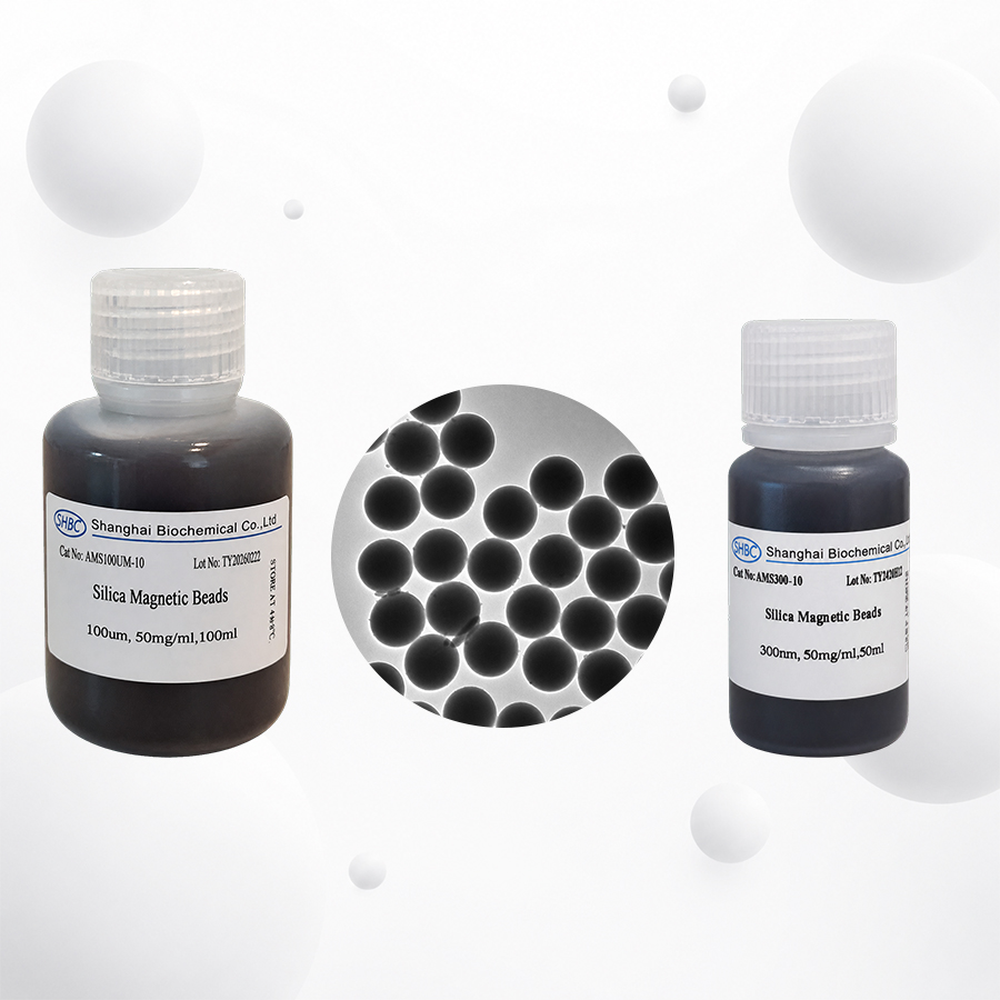 How Silica Magnetic Beads Work: Quick AnswerSilica magnetic beads extract DNA and RNA through a reversible bind–wash–elute process.First, a lysis buffer breaks open cells, viruses or other biological materials and releases nucleic acids into solution. A binding buffer containing salts—and often alco
-
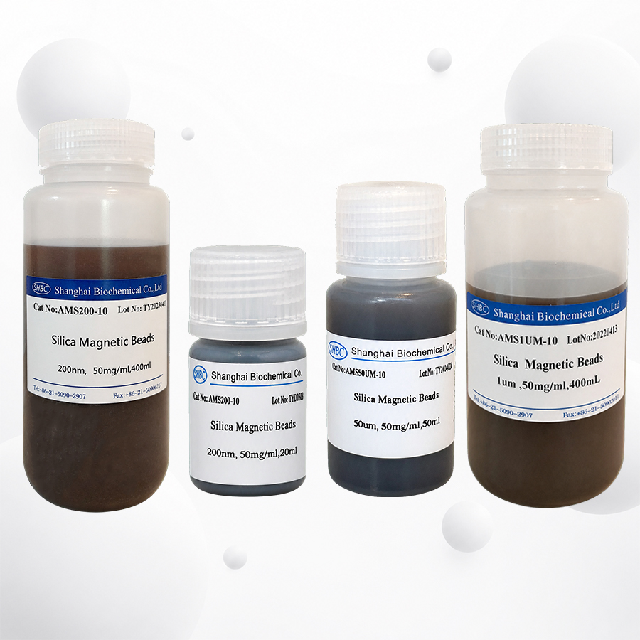 Overview of the Silica Magnetic Bead MarketSilica magnetic beads are functional magnetic particles used to extract and purify DNA or RNA from biological, clinical, food, environmental and research samples.A typical bead contains a magnetic core and a silica or silica-like outer surface. Under suitab
-
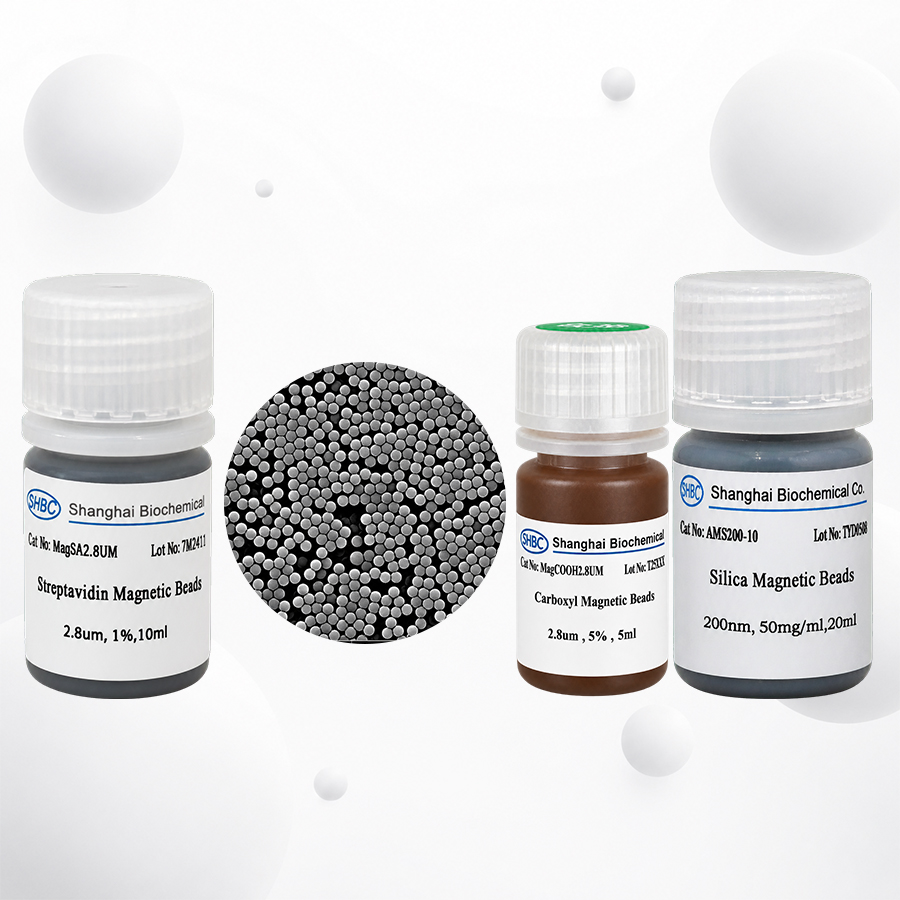 How to Choose Silica Magnetic Beads: Quick AnswerChoose silica magnetic beads according to the complete extraction workflow—not according to particle diameter or catalog price alone.The correct bead must match:The target nucleic acid;The sample matrix;The expected nucleic acid concentration;The requ
-
 Quick Answer: Top 10 Microsphere Manufacturers Serving EuropeThe top 10 microsphere manufacturers and specialist particle suppliers serving European lateral flow assay developers in 2026 are:Merck EstaporSHBCBangs LaboratoriesPolysciencesThermo Fisher ScientificJSR Life SciencesMagsphereSpherotechmi


