


Polystyrene Microspheres (PS Microspheres): Properties, Types and Applications
Polystyrene microspheres are spherical polymer particles manufactured from styrene or styrene-based copolymers. They are available in particle sizes ranging from the nanometer scale to tens or hundreds of micrometers and can be supplied as aqueous suspensions or dry powders.
When dispersed in water, these particles are often referred to as polystyrene latex beads. However, “latex microspheres” is a broader term that can also describe particles made from other polymers. The commercial term “PS microspheres” may include both submicron polystyrene particles and true micron-sized spheres.
Polystyrene microspheres are valued for their controllable particle size, relatively low density, high refractive index, adaptable surface chemistry and compatibility with optical detection methods. Plain particles can adsorb proteins through hydrophobic interactions, while functionalized particles support covalent attachment of antibodies, proteins, peptides and other biomolecules.
Their controllable particle size, optical properties and adaptable surface chemistry make them useful in diagnostics, instrument calibration, particle research and materials development.
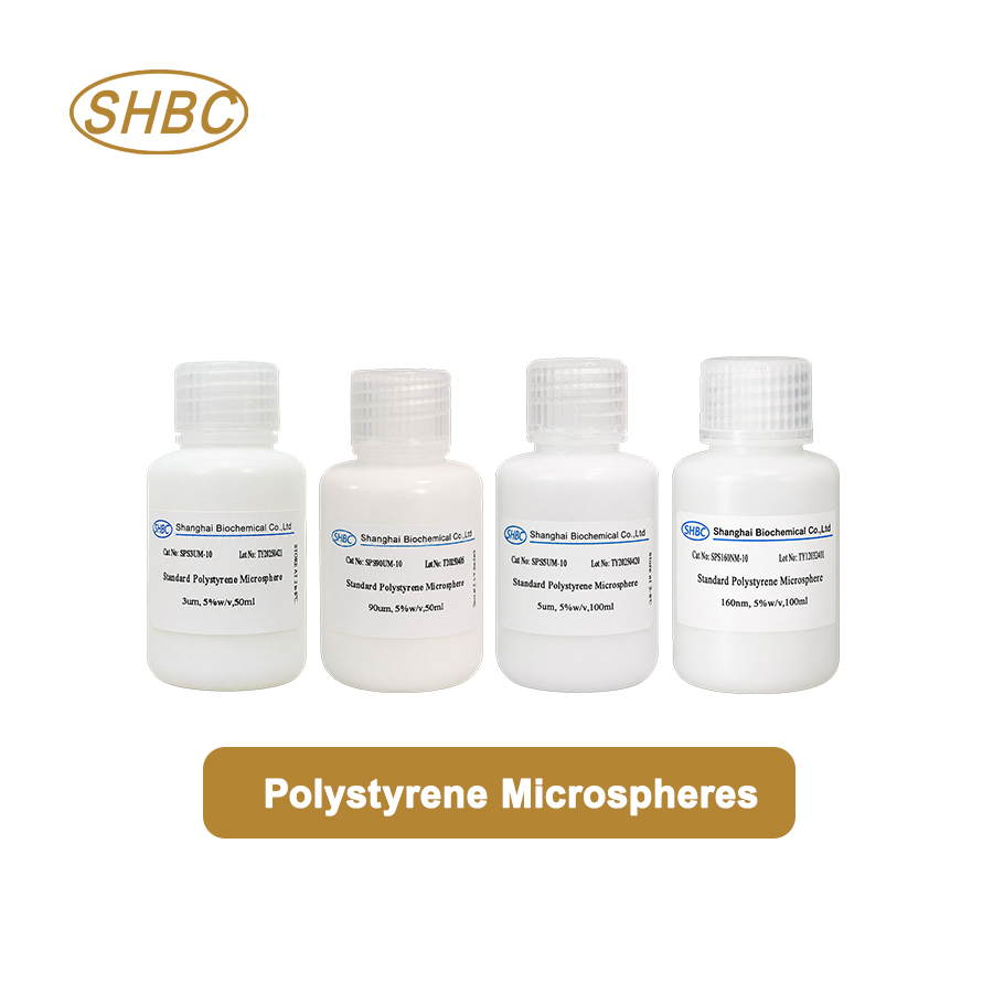
What Are Polystyrene Microspheres?
Polystyrene microspheres consist primarily of polymerized styrene arranged into spherical particles. Depending on the manufacturing method, they can be produced with different particle sizes, size distributions, surface charges, degrees of crosslinking and functional groups.
Typical polystyrene has a density of approximately 1.05 g/cm³ and a refractive index of about 1.59 in the visible wavelength range. Actual values can vary with particle formulation, crosslinking, dyes and incorporated functional materials.
The aromatic polymer surface is naturally hydrophobic. This characteristic allows plain polystyrene particles to adsorb many proteins without requiring a chemical coupling reaction. However, adsorption strength, protein orientation and long-term stability depend on the protein, pH, ionic strength, surfactant concentration and blocking conditions.
Polystyrene microspheres can also be manufactured with reactive surface groups such as carboxyl or amino groups. These functionalized particles provide more controlled routes for immobilizing antibodies and other ligands.
Types of Polystyrene Microspheres
Selecting the appropriate type of PS microsphere begins with understanding the difference between plain, functionalized and optically modified particles.
Plain Polystyrene Microspheres
Plain PS microspheres have a hydrophobic polymer surface without a deliberately introduced reactive functional group.
They are commonly used for:
Passive adsorption of antibodies and proteins
Particle size and instrument calibration
Microscopy and imaging studies
Colloid and surface-interaction research
Filtration and particle-retention testing
Model particle experiments
Passive adsorption is relatively simple and does not require a chemical activation step. However, the amount and orientation of adsorbed protein may be more difficult to control than with covalent coupling.
Carboxyl Polystyrene Microspheres
Carboxyl polystyrene microspheres contain surface carboxyl groups. These groups provide a negative surface charge under suitable pH conditions and can be activated using carbodiimide chemistry.
EDC, often used together with NHS or sulfo-NHS, activates the carboxyl groups so they can react with primary amines on antibodies, proteins or peptides. This produces a stable amide bond between the particle and the biomolecule.
Carboxyl-functionalized particles are widely selected when stronger attachment and better resistance to washing are required.
Amino Polystyrene Microspheres
Amino-functionalized particles contain primary amine groups on their surfaces. These groups can react with NHS esters, aldehydes, activated carboxyl groups and other amine-reactive compounds.
Amino PS microspheres are suitable for further surface modification and for coupling molecules that contain compatible reactive groups. The coupling method must be selected carefully because antibodies and proteins also contain multiple amines.
Colored and Fluorescent Polystyrene Microspheres
Colored PS microspheres contain pigments or dyes that provide a visible signal. Fluorescent PS microspheres incorporate fluorophores for excitation and emission at selected wavelengths.
They may be used in:
Lateral flow assays
Flow cytometry
Fluorescence microscopy
Cell counting
Multiplex assays
Optical instrument calibration
Tracer and imaging experiments
When choosing fluorescent particles, excitation wavelength, emission wavelength, fluorescence intensity, photostability and compatibility with the instrument’s filters should all be considered.
How Are Polystyrene Microspheres Manufactured?
Polystyrene microspheres can be manufactured using emulsion polymerization, dispersion polymerization, suspension polymerization or seeded-growth methods.
Emulsion Polymerization
Emulsion polymerization is commonly used to produce nanometer-scale and submicron polystyrene particles. Styrene monomer is dispersed in an aqueous phase containing an initiator and, depending on the formulation, a surfactant or stabilizer.
The concentrations of monomer, initiator, surfactant and electrolyte influence particle nucleation, growth and final particle size.
Dispersion Polymerization
Dispersion polymerization is often used to prepare relatively uniform micron-sized particles. Polymerization begins in a medium in which the monomer is soluble but the growing polymer becomes insoluble.
A stabilizer prevents the newly formed particles from uncontrolled aggregation. Reaction conditions can be adjusted to control particle size and size distribution.
Suspension Polymerization
Suspension polymerization is generally more suitable for producing larger particles. Monomer droplets are suspended in a continuous liquid phase and polymerized inside the droplets.
Particle size is influenced by mixing speed, stabilizer concentration, viscosity and droplet formation. Compared with carefully controlled emulsion or dispersion processes, suspension polymerization may produce a broader particle size distribution.
Seeded Particle Growth
In a seeded-growth process, existing particles serve as seeds for additional polymer formation. This method can increase particle diameter while maintaining better control over particle uniformity.
Following polymerization, the particles may undergo washing, concentration adjustment, classification, surface functionalization, dye incorporation and quality testing.
Key Properties of PS Microspheres
The performance of polystyrene microspheres depends on more than their nominal particle diameter. Several physical and chemical properties should be evaluated together.
Particle Size
Particle size affects sedimentation, Brownian motion, surface area, optical scattering, assay kinetics and interaction with biological samples.
Smaller particles provide a larger surface area per unit mass. They may increase binding capacity and remain suspended for longer periods, but they can be more difficult to separate or observe directly.
Larger particles are generally easier to visualize and separate. However, they settle more rapidly and may require gentle resuspension before sampling.
Particle Size Distribution
A narrow particle size distribution improves consistency between experiments. It is especially important in calibration, flow cytometry, quantitative imaging and assays where particle size affects signal intensity.
Particle uniformity is often expressed using the coefficient of variation:
CV = standard deviation ÷ mean particle diameter × 100%
A lower CV indicates a narrower size distribution. The acceptable CV depends on the application and measurement method.
Surface Area
Smaller particles have a higher surface-area-to-volume ratio than larger particles. This can increase the number of available sites for protein adsorption or surface functionalization.
However, a higher theoretical surface area does not automatically guarantee better assay performance. Steric hindrance, ligand orientation, surface charge, blocking and mass transfer can all affect the usable binding capacity.
Surface Charge
Surface charge influences colloidal stability and interactions with proteins, cells, membranes and other particles.
Zeta potential is frequently used to evaluate the electrostatic stability of a particle suspension. A high absolute zeta potential can help particles repel each other, although steric stabilizers and surfactants may also provide stability when the zeta potential is relatively low.
Because zeta potential depends on pH, ionic strength and buffer composition, results should always be interpreted under clearly defined measurement conditions.
Refractive Index
Polystyrene has a relatively high refractive index, which produces strong light scattering compared with many aqueous media. This makes PS particles useful in optical detection, microscopy and instrument calibration.
Signal intensity still depends on particle size, wavelength, detector configuration and the refractive index of the surrounding medium.
Solid Content
PS microsphere suspensions are commonly supplied at a defined weight or volume concentration. Solid content affects the number of particles added to an assay and must be considered when comparing products.
Two suspensions with the same weight percentage may contain very different particle numbers if their particle diameters differ. Particle number concentration should therefore be calculated when the application requires a controlled bead-to-analyte ratio.
Chemical and Thermal Compatibility
Polystyrene is compatible with water and many mild aqueous buffers but can swell or dissolve in certain organic solvents. Aromatic solvents, chlorinated solvents and some ketones may damage the particles.
Temperature, solvent exposure and sterilization methods should be validated before use. Standard PS particles should not be assumed to tolerate autoclaving or aggressive organic solvents.
How to Choose Polystyrene Microspheres
The correct PS microsphere should be selected according to the application rather than particle size alone.
1. Define the Intended Application
First determine whether the particles will be used for protein immobilization, calibration, fluorescence detection, filtration testing, microscopy or materials research.
An assay-development project may prioritize surface chemistry and protein-binding performance, while an instrument-calibration project may require a highly uniform diameter and stable optical signal.
2. Select the Particle Size
Particle diameter influences surface area, sedimentation rate, reaction kinetics and detection method.
Submicron particles are suitable when a large surface area and stable suspension are important. Micron-sized particles are often preferred when direct visualization, flow analysis or easier separation is required.
The sample matrix and instrument detection range should also be considered.
3. Choose the Surface Chemistry
Plain PS microspheres can be used for passive adsorption of proteins. Carboxyl particles support EDC/NHS-mediated covalent coupling, while amino particles provide alternative conjugation routes.
Covalent coupling is generally preferred when the immobilized biomolecule must remain attached during repeated washing, storage or exposure to complex samples.
4. Review Particle Uniformity
Check the mean particle size, particle size distribution and CV. Highly uniform particles are especially important for quantitative optical measurements and calibration.
The measurement method should also be reviewed. DLS measures hydrodynamic diameter and can be strongly influenced by aggregates, while electron microscopy measures particles in a dry state. Results from different methods may therefore not be identical.
5. Confirm the Dispersion Medium
The suspension may contain water, buffer, surfactant, stabilizer or preservative. These components can affect antibody adsorption, enzyme activity, cell compatibility and downstream chemical reactions.
If the supplied medium is incompatible with the application, a validated washing or buffer-exchange procedure may be required.
6. Evaluate Optical Requirements
For colored or fluorescent particles, verify the absorption, excitation and emission characteristics. The signal must be compatible with the intended reader, microscope or flow cytometer.
Potential fluorescence leakage, photobleaching and signal variation between batches should also be evaluated.
7. Check Packaging and Batch Requirements
Development work may require only a small volume, while commercial manufacturing may require consistent large batches.
Particle-size specifications, solid content, surface functional-group density, packaging volume and batch-to-batch consistency should be discussed before scale-up.
Particle Size and Morphology Characterization
No single analytical method provides all the information needed to characterize PS microspheres.
Dynamic light scattering is useful for measuring the hydrodynamic size of submicron particles in suspension. However, a small amount of aggregation can cause a disproportionately large increase in the reported diameter.
Laser diffraction can cover a broader particle size range but may have lower resolution for distinguishing closely spaced particle populations.
Optical microscopy is convenient for inspecting sufficiently large particles and identifying visible aggregation or contamination.
Scanning electron microscopy provides direct information about particle shape, surface appearance and dry-state diameter. Sample preparation must be controlled because drying can bring particles together and create apparent aggregates that were not present in the original suspension.
SEM imaging can confirm particle morphology, surface appearance and the presence of fused, deformed or aggregated particles.
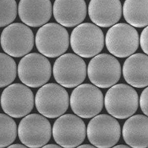
For critical applications, particle size data should be interpreted together with microscopy, solid-content measurements and application-specific performance tests.
Protein and Antibody Immobilization
Polystyrene microspheres are frequently used as solid supports for antibodies, antigens and other proteins.
Passive Adsorption on Plain PS Microspheres
Plain PS microspheres can adsorb proteins through hydrophobic and electrostatic interactions. A typical development process includes:
Resuspending the particles completely.
Adding the protein under a controlled pH and ionic strength.
Incubating for a defined time.
Blocking the remaining nonspecific adsorption sites.
Washing away unbound material.
Resuspending the coated particles in a suitable storage buffer.
The optimal protein concentration should be determined experimentally. Excess protein does not always improve performance and may increase nonspecific binding or aggregation.
Passive adsorption is convenient, but the protein can attach in different orientations. Some antigen-binding sites may face the particle surface and become less accessible.
Covalent Coupling to Carboxyl PS Microspheres
Carboxyl PS microspheres can be activated with EDC and NHS or sulfo-NHS. The activated groups then react with primary amines on the antibody or protein.
Important variables include:
Activation-buffer composition
Activation pH
EDC and NHS concentration
Reaction time
Antibody concentration
Coupling-buffer composition
Blocking reagent
Washing conditions
Final storage buffer
Covalent coupling can improve attachment stability, but it does not automatically ensure correct antibody orientation. Coupling efficiency and retained biological activity should both be measured.
Applications of Polystyrene Microspheres
Immunoassays and Latex Agglutination
Antibodies or antigens can be immobilized on PS particles to produce visible or instrument-detected agglutination. Particle size, surface chemistry, coating density and blocking conditions affect sensitivity and background.
Plain PS particles may be suitable for passive coating, while carboxyl-functionalized particles are preferred when stable covalent coupling is required.
Instrument Calibration
Uniform PS microspheres can be used to evaluate particle detection, optical alignment, counting precision and size response.
A general-purpose PS microsphere should not automatically be described as a certified size standard. Applications requiring traceability should use products supplied with the appropriate certification and measurement documentation.
Flow Cytometry and Cell Counting
Fluorescent PS microspheres can support detector setup, fluorescence compensation, particle counting and performance monitoring.
The particle diameter and fluorescence intensity must fall within the instrument’s detection range. Aggregation should be minimized because bead doublets and clusters can appear as larger particle populations.
Microscopy and Imaging
PS microspheres provide particles with controlled sizes and optical properties for microscopy evaluation, image-analysis development and resolution testing.
Colored or fluorescent particles can improve contrast and support multi-channel imaging studies.
Lateral Flow and Biosensors
Colored and fluorescent PS microspheres can be functionalized with antibodies or other recognition molecules and used as detectable labels.
Surface chemistry, signal intensity, membrane migration, conjugate stability and nonspecific interactions must be optimized as a complete system.
Filtration and Particle-Retention Testing
Particles with controlled diameters can be used to study filter retention, membrane pore behavior and particle transport.
The selected bead size should be appropriate for the expected pore size and analytical detection method.
Colloid and Materials Research
PS microspheres are frequently used as model particles in studies of aggregation, sedimentation, self-assembly, surface interactions and photonic structures.
Because standard polystyrene is not biodegradable, its suitability for biomedical or environmental applications should be evaluated according to the intended use.
Handling and Storage
Correct handling helps preserve particle concentration, uniformity and surface performance.
Follow the storage temperature specified by the supplier.
Do not freeze the suspension unless freeze stability has been confirmed.
Keep the container tightly closed to prevent evaporation and contamination.
Avoid allowing the suspension to dry around the bottle neck or cap.
Gently resuspend the particles before withdrawing a sample.
Use clean pipette tips and do not return removed material to the original container.
Protect fluorescent particles from prolonged light exposure.
Avoid exposing the particles to incompatible solvents or extreme pH.
Record the lot number, opening date and storage history.
Large particles may sediment during storage without being irreversibly aggregated. Sedimentation alone does not necessarily indicate that the product has failed.
How to Resuspend Settled PS Microspheres
Begin by gently inverting or rolling the closed container. If necessary, use brief vortex mixing at a controlled speed.
Sonication can help disperse some particle suspensions, but excessive sonication may heat the sample, damage surface coatings or affect biomolecules already attached to the particles. It should only be used under validated conditions.
After resuspension, inspect the sample using an appropriate method such as optical microscopy, DLS or flow cytometry.
Do not automatically filter, centrifuge or readjust the solid content of an aggregated suspension. These operations can remove part of the particle population and change both particle-size distribution and concentration.
If irreversible aggregates remain, isolate the affected material and contact the supplier before using it in a sensitive assay or calibration procedure.
Common Causes of Aggregation
PS microsphere aggregation may result from:
Freezing or overheating
Drying at the container opening
High salt concentration
Incompatible buffer composition
Incorrect pH
Removal or dilution of stabilizing surfactants
Protein overloading
Insufficient blocking
Repeated contamination
Incompatible organic solvents
Excessive centrifugation
Long-term storage under unsuitable conditions
When aggregation occurs after a buffer change or biomolecule-coupling step, investigate the formulation and process conditions before assuming that the original particles were defective.
Quality-Control Parameters
Important quality-control parameters for polystyrene microspheres may include:
Mean particle diameter
Particle size distribution and CV
Particle morphology
Solid content
Particle number concentration
Surface charge or zeta potential
Surface functional-group density
Dispersion stability
Color or fluorescence intensity
Residual surfactant or preservative
Protein-coupling capacity
Functional assay performance
Batch-to-batch consistency
The most relevant tests depend on the application. A particle that meets a physical size specification may still require biological or optical performance testing before use in a finished diagnostic product.
Polystyrene vs PMMA and Silica Microspheres
Polystyrene, PMMA and silica microspheres have different material properties and should not be treated as interchangeable.
Polystyrene has a relatively high refractive index and hydrophobic surface. It is particularly useful for optical applications, passive protein adsorption and polymer-particle research.
PMMA generally has a lower refractive index and a higher density than polystyrene. Its solvent resistance and mechanical behavior also differ from PS.
Silica microspheres are denser, more rigid and generally more hydrophilic. Their surfaces can be modified through silane chemistry, but they sediment faster than similarly sized PS particles.
The best material depends on the required density, refractive index, surface chemistry, solvent resistance and detection method.
Frequently Asked Questions
Are polystyrene microspheres the same as latex beads?
Polystyrene microspheres dispersed in water are often called polystyrene latex beads. However, latex is a general term for a colloidal polymer dispersion and does not always mean that the particles are made from polystyrene.
Can plain PS microspheres bind antibodies?
Yes. Plain PS microspheres can adsorb many antibodies through hydrophobic and electrostatic interactions. The adsorption conditions and blocking method must be optimized to preserve antibody activity and control nonspecific binding.
Should I choose plain or carboxyl PS microspheres?
Choose plain PS microspheres when simple passive protein adsorption is sufficient. Choose carboxyl PS microspheres when stable covalent coupling and stronger resistance to washing or storage are required.
What particle size should I use?
The answer depends on the detection method, required surface area, sedimentation behavior and sample matrix. Smaller particles provide more surface area per unit mass, while larger particles are generally easier to visualize and separate.
Why do PS microspheres settle?
Micron-sized particles can settle under gravity, especially during long-term storage. Normal sedimentation is usually reversible with gentle mixing. Irreversible clumps that remain after controlled resuspension may indicate aggregation.
Can PS microspheres be frozen?
Freezing is generally not recommended unless the product has been specifically validated for freeze-thaw stability. Ice formation can concentrate salts and particles, leading to irreversible aggregation.
Can polystyrene microspheres be autoclaved?
Standard PS microspheres should not be assumed to tolerate autoclaving. High temperature can change the particle surface, dispersion stability and particle shape. Use a validated sterilization method appropriate for the specific product.
Why can DLS and SEM report different particle sizes?
DLS measures hydrodynamic diameter in suspension, including the effect of the surrounding liquid and surface layers. SEM measures dry particles directly. Sample preparation, aggregation and the measurement principle can therefore produce different results.
Are polystyrene microspheres biodegradable?
Conventional polystyrene is not considered biodegradable. PS microspheres are widely used as research, diagnostic and industrial particles, but applications requiring biodegradable materials should consider another polymer.
Should PS microspheres be supplied as a powder or suspension?
A suspension is generally easier to resuspend consistently and reduces airborne-particle exposure. Powder may be useful when a custom solvent or high solid content is required, but complete redispersion can be more difficult.
Conclusion
Polystyrene microspheres provide a versatile platform for instrument calibration, immunoassays, microscopy, filtration studies, optical detection and materials research. Reliable performance depends on selecting the correct particle size, size distribution, surface chemistry, solid content and dispersion medium.
Plain PS microspheres support passive protein adsorption and general particle applications, while carboxyl-functionalized particles provide a practical route for covalent antibody and protein immobilization. Colored and fluorescent variants can support visual or optical detection.
SANYU GROUP supplies Polystyrene Microspheres in standard nano- and micron-scale particle sizes for assay development, calibration, particle research and materials applications. Carboxyl Polystyrene Microspheres are also available for projects requiring EDC/NHS-mediated biomolecule coupling.
For applications requiring a nonstandard particle size, surface group, solid content, color, fluorescence, dispersion medium or packaging format, SANYU GROUP supports custom particle development, OEM/ODM cooperation and scale-up manufacturing.
Contact our technical team with your required particle size, surface chemistry, solid content, application and estimated volume to discuss an appropriate PS microsphere specification.
Related Blogs
-
 CD3 CD28 magnetic beads have revolutionized T cell activation, offering a highly efficient and scalable solution for researchers and clinicians working in immunology and cell therapy. These antibody-coated magnetic beads mimic natural antigen-presenting cells, providing controlled and reproducible s
 -
 Latex microspheres, also known as polystyrene microspheres, PS microspheres, etc. Polystyrene microspheres are a general term for polymer microspheres formed by self polymerization of styrene or copolymerization of styrene with other monomers with specific functional groups. The surface of polystyre
 -
 NHS activated magnetic beads are revolutionizing bioconjugation techniques in molecular biology and biotechnology. These powerful tools enable researchers to efficiently attach proteins, antibodies, peptides, and nucleic acids, significantly enhancing purification and analysis processes. Understandi
 - Introduction to Streptavidin Magnetic BeadsStreptavidin magnetic beads (SA beads for short) refer to magnetic microspheres with specific bioactive substances (streptavidin) modified on their surface.Streptavidin magnetic beads are a type of biological nanomagnetic beads with a surface functional gro

