


How to Choose Silica Magnetic Beads for DNA and RNA Extraction
Choose silica magnetic beads by testing how the particles perform with your sample, reagents and magnetic platform. Particle diameter is useful for building a shortlist, but it cannot predict extraction recovery, inhibitor removal or reliable automation by itself.
A suitable bead must provide accessible binding surface, consistent dispensing, effective magnetic collection and adequate release of DNA or RNA during elution. These requirements need to be assessed together.
Begin by defining the target nucleic acid, sample matrix, input amount, required fragment range and downstream assay. Then compare candidate bead grades using a controlled extraction method. Select the bead and buffer combination that meets the application requirements across representative samples.
Define the Extraction Requirements First
A useful selection brief describes the extraction task before specifying a diameter. Include:
Target nucleic acid: genomic DNA, total RNA, viral nucleic acid, cfDNA or small RNA.
Sample matrix and preparation: blood, plasma, swabs, cells, tissue, plants or other material.
Sample input and expected target abundance.
Required fragment-size range and integrity.
Manual rack or automated platform, including vessel format.
Binding and wash chemistry, elution volume and downstream assay.
These details identify the main constraints. High-input tissue DNA can challenge capacity and mixing, while low-abundance plasma DNA requires efficient recovery of the relevant fragments. RNA workflows also need suitable stabilization and RNase control.
The sample matrix determines which contaminants and handling problems must be addressed. Plant lysates, viscous saliva and plasma should not be represented only by purified DNA in water during final evaluation.
It also helps to distinguish raw-sample extraction from cleanup of an existing nucleic acid preparation. A successful cleanup test shows useful binding and elution behavior, but it does not demonstrate adequate lysis or inhibitor removal from the original biological sample.
A Practical Selection Framework
Use three stages to organize the decision:
Define the application and measurable acceptance criteria.
Shortlist particles by surface type, size information, suspension specification and magnetic handling.
Compare complete extraction performance, then confirm the selected method across samples and lots.
This approach separates material specifications from application results. A size distribution describes the particles, while a recovery result describes a particular extraction experiment. Both are useful, but they answer different questions.
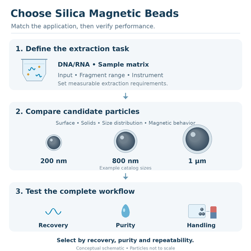
The schematic summarizes the selection process: define the sample and target, compare particle candidates, and test recovery, purity and handling in the intended workflow. The illustrated diameters are example catalog sizes, not a size-to-application chart or a prediction of extraction performance.
Particle Size and Size Distribution
Interpret nominal diameter in context
Particle size affects geometric surface area, settling, fluid resistance and handling. For nonporous spheres of comparable composition and density, smaller particles provide more geometric surface area per unit mass.
Actual nucleic acid capacity also depends on accessible silica, surface structure and the solution conditions. A smaller nominal diameter therefore does not automatically mean higher usable capacity or better recovery.
Magnetic collection depends on the particle's magnetic properties as well as its size and the surrounding liquid. Larger particles are not universally faster, and smaller particles are not automatically unsuitable for automated extraction.
Choose a practical range of candidates, then assess dispersion, capture and elution on the intended platform.
Ask how size was measured
SEM or TEM can describe particle dimensions in prepared specimens. Dynamic light scattering measures hydrodynamic behavior in suspension. Its result can be affected by hydration and aggregation and should not be treated as an identical measurement of dry particle diameter.
For each size specification, ask for the measurement method, sample preparation and reported statistic. Also establish whether a distribution is expressed on a number, volume or intensity basis.
A diameter from one method can differ from another without proving that either result is incorrect.
Keep CV and DLS PDI distinct
A particle-size coefficient of variation describes standard deviation relative to mean diameter for the specified distribution. DLS polydispersity index comes from a different analysis of the scattering correlation data.
Do not compare a microscopy-based CV directly with a DLS PDI as though they were interchangeable acceptance limits. A universal CV or PDI threshold also cannot establish extraction performance.
Use consistent measurement methods when comparing lots, and combine physical characterization with a functional extraction test.
Surface Chemistry and Buffer Compatibility
The accessible silica surface provides the nucleic acid-binding interface. Standard silanol-rich silica, carboxyl-modified silica and amino-modified particles should be identified clearly rather than grouped together under one magnetic-bead label.
For ordinary silica extraction, binding is reversible and depends on the surrounding solution. Many methods use chaotropic salts and alcohol during binding, followed by compatible washes and low-salt elution. Buffer composition influences both retention and release.
Confirm the supplied suspension medium and relevant additives. Replacing one bead stock with another can change the final reagent mixture, particularly when stock volumes differ.
Screen the candidate in the intended binding and wash conditions. Look for aggregation, poor resuspension, target loss during washing and incomplete elution. A result obtained in one buffer does not establish compatibility with every silica extraction formulation.
SPRI-type methods commonly use carboxyl-functionalized particles with PEG and salt. Their cleanup or fragment-selection ratios should not be transferred automatically to ordinary silica-coated extraction beads.
Surface area and coating descriptions can support a shortlist. The final decision should include recovery and contaminant removal under the actual extraction conditions.
Magnetic Capture and Redispersion
Measure capture in the working liquid
Evaluate magnetic collection using the intended magnet, vessel, liquid volume and sample mixture. Collection in water is useful for an initial handling check, but it does not reproduce every extraction environment.
Record whether particles collect adequately before liquid removal, where the bead mass forms and whether material remains suspended. Sample viscosity and vessel geometry can change the result.
Visible clarity alone is not a complete measure of bead retention. Where carryover matters, use an appropriate measurement or a functional check of the transferred fraction.
Incomplete capture can lose bead-bound nucleic acid during aspiration. A short advertised collection time is therefore meaningful only when its test conditions are specified.
Check repeated resuspension
Particles must make effective contact with nucleic acid during binding and with the liquid during washing and elution. A bead that collects readily but forms persistent clumps can create a different handling problem.
Check resuspension after each stage, including the final wash and any prescribed drying step. Evaluate the complete sequence because behavior can change after exposure to sample proteins or extraction reagents.
Superparamagnetic behavior supports repeated magnetic manipulation but does not guarantee freedom from aggregation.
Do not assume that increasing magnetic content or extending collection time will correct inadequate mixing. Identify whether the limiting step is dispersion, collection, aspiration or release.
Binding Capacity, Solids Concentration and Bead Dose
Binding capacity should specify the nucleic acid, fragment characteristics, buffer conditions, bead quantity and measurement method. A value reported per milliliter of suspension is difficult to compare unless the suspension's solids concentration is also known.
Distinguish the amount adsorbed during binding from the amount recovered after washing and elution. High adsorption with poor release can still produce an unsuitable extraction result.
Equal suspension volumes do not necessarily contain equal particle masses. By definition, a suspension specified as 5% w/v contains 50 mg/mL of solids. A 20 μL aliquot of that suspension contains 1 mg of solids. This arithmetic helps compare quantities; it does not establish an optimal extraction dose.
Use a defined dose series when developing the method. Too little accessible surface can limit capture, while additional particles may increase handling demands or leave more material near the final aspiration point.
Compare recovered target, purity and carryover as the dose changes. Select the dose that meets the workflow requirements, rather than assuming that the largest addition is best.
For high-input samples, evaluate the working range before saturation. For low-input samples, focus on efficient recovery and transfer losses as well as nominal capacity.
Selection Priorities for Different Nucleic Acid Targets
Target | Main selection priority | Useful evaluation |
|---|---|---|
Routine genomic DNA | Adequate capacity and inhibitor removal | Recovered mass, amplification and fragment distribution |
High-molecular-weight DNA | Preservation and release of long molecules | Size-sensitive integrity analysis and downstream suitability |
Total RNA | RNase control, recovery and acceptable DNA contamination | RNA integrity, target recovery and relevant no-RT controls |
Viral DNA or RNA | Low-target recovery in the collection medium | Extraction and amplification controls across representative samples |
Cell-free DNA | Recovery of the required short-fragment range | Sensitive quantification combined with fragment analysis |
Small RNA | Retention and elution of the desired short species | Size-sensitive or target-specific recovery measurements |
DNA and high-molecular-weight DNA
Routine PCR compatibility does not establish suitability for applications requiring long DNA molecules. For high-molecular-weight DNA, evaluate lysis, mixing, transfer and elution alongside the bead.
Gentle handling can preserve length, but insufficient mixing can reduce extraction efficiency. The selected method must balance those needs and be assessed with a suitable integrity measurement.
RNA and small RNA
Silica can bind DNA and RNA, so the bead surface alone does not guarantee selective purification of one species. Enzyme treatment and buffer conditions may be needed to manage unwanted nucleic acid.
RNA integrity depends on sample preservation and RNase control throughout processing. A bead cannot reverse degradation that has already occurred.
Small RNA needs specific evaluation. Good recovery of longer RNA does not demonstrate retention of microRNA or other short species. Confirm the required range with relevant controls.
Viral nucleic acids and cfDNA
For scarce targets, evaluate recovery at the intended low input rather than relying only on a high-load DNA standard. Include the actual collection medium or representative plasma matrix.
For cfDNA, total DNA concentration can include contaminating cellular DNA. Combine sensitive quantification with fragment analysis and target-relevant measurements where needed.
Smaller elution volumes can increase concentration, but the recovered mass and usable volume still matter. Assess the complete result rather than concentration alone.
Automation and Scale-Up
Liquid handlers and magnetic-rod systems manipulate beads differently. Liquid handlers commonly retain particles while transferring liquids; rod systems move particles between reagent wells and must release them effectively for mixing.
A bead should be evaluated on the specific instrument and consumables, with appropriate working volumes. Compatibility with one magnetic rack does not demonstrate compatibility with every automated extractor.
During scale-up, assess stock mixing, dispensing consistency, settling during pauses, bead release and aspiration behavior. Check whether early and late samples receive comparable particle quantities.
Compare the automated method with an established manual reference where practical. This can help distinguish a chemistry limitation from a liquid-handling or mixing problem.
Include representative plate positions and processing conditions in confirmation runs. Filling or extraction at production scale can expose variation that was not apparent in a few manually prepared tubes.
A Comparative Evaluation Plan
Set acceptance criteria before screening
Define the required target recovery, integrity, downstream assay performance, processing time and allowable carryover. Criteria should reflect the application rather than a generic supplier specification.
Include negative extraction controls and representative difficult samples. Low-abundance material is particularly useful when the intended workflow must recover scarce targets.
Decide what the comparison is testing
A fixed-buffer comparison helps assess how candidate beads behave under one defined formulation. Comparing individually optimized methods answers a different question: which complete bead and buffer system works best.
State which comparison is being made. Keep sample input, control material, elution conditions and analytical measurements consistent where needed for interpretation.
When comparing bead quantities, use the stated solids concentration and document the dose. Matching particle mass can provide a useful starting point, but different particles may require different optimized doses.
Evaluate several outcomes together
Outcome | What to record |
|---|---|
Recovery | Target quantity and recovered eluate volume |
Purity and inhibition | Appropriate purity information and downstream inhibition controls |
Integrity | Fragment distribution or RNA integrity when relevant |
Handling | Collection, resuspension, bead loss and carryover |
Repeatability | Variation across samples, runs, positions and lots |
A fluorescence assay provides useful nucleic acid quantification but is not itself a purity test. UV measurements offer additional information, although contaminants and mixed nucleic acids can complicate interpretation.
Likewise, a lower Ct does not by itself prove greater binding capacity. Extraction input, concentration, inhibition and assay input all affect the comparison.
Confirm the selected method
After initial screening, refine dose, mixing and other supported conditions for the shortlisted candidate. Document the final formulation and instrument settings.
Repeat the method across the relevant input range and sample types, then evaluate additional bead lots. Preserve a reference method or control material for future comparisons.
A purified nucleic acid spike helps test some recovery steps, but it does not necessarily reproduce release from cells or viral particles. Include material that challenges the required sample preparation when lysis is part of the application.
Documentation, Supply and Cost per Successful Extraction
Ask for a specification that clearly identifies the surface, size measurement, solids concentration, supplied medium, storage conditions and shelf life. Review a lot-specific COA where available and distinguish measured results from nominal specifications.
For recurring or OEM supply, discuss packaging, batch volumes, traceability and notification of changes that could affect the method. Specify any required nuclease or other background testing and agree on how it will be assessed.
Use application-relevant acceptance tests for incoming material. Matching a nominal diameter alone is insufficient when the extraction process depends on collection and release behavior.
Compare cost at the validated working dose. Include reagents, processing time, repeat extractions and failed downstream analyses where they materially affect the project.
The lowest price per milliliter may not produce the lowest cost per usable extraction. Conversely, a premium specification has limited value if it does not improve a requirement that matters to the application.
SANYU Silica Magnetic Bead Options
SANYU manufactures functional particles, including silica magnetic beads for nucleic acid extraction development. Its published catalog includes particle listings such as 200 nm, 300 nm, 800 nm and 1 μm, alongside other sizes and surface-modified grades.
These sizes are candidate options rather than fixed recommendations for particular sample types. Confirm the surface chemistry and specification of the selected grade before evaluation.
For a sample request, provide the target nucleic acid, matrix, input volume, extraction platform, buffer approach and downstream assay. Include the required fragment range and any limits on processing time or elution volume.
Particle size, suspension concentration and surface customization can be discussed against those requirements. Comparative testing then establishes which candidate and formulation meet the extraction targets.
Frequently Asked Questions
What is the best silica magnetic bead size for DNA or RNA extraction?
There is no universal best diameter. Select a particle by evaluating surface chemistry, capture, resuspension and extraction performance with the intended sample, buffers and magnetic platform.
Are smaller silica magnetic beads always better?
No. Smaller particles can provide more geometric surface area under comparable conditions, but usable capacity, magnetic handling, aggregation and elution also affect recovery. Nominal size alone cannot determine the result.
Can the same silica magnetic beads extract both DNA and RNA?
Some silica beads can support both workflows with suitable reagents. DNA and RNA methods may still require different preparation, enzyme treatments and recovery checks, especially when small RNA or long DNA matters.
Does a 5% bead suspension always mean 50 mg/mL?
Only when 5% is specified as w/v. That definition corresponds to 50 mg/mL. A percentage without a stated basis should be clarified before calculating the bead mass or comparing doses.
Does the highest binding capacity identify the best bead?
No. Capacity must be interpreted with its test conditions. Recovery after washing and elution, inhibitor removal, integrity and reliable handling are also necessary for a useful extraction.
Can beads be substituted into an existing extraction kit?
Treat substitution as a method change requiring evaluation. Surface chemistry, stock medium, solids concentration and capture behavior may differ, even when the replacement has the same nominal diameter.
Is a datasheet enough to approve a bead for automated extraction?
A datasheet supports screening. Confirm the selected grade using the intended instrument, consumables, sample range and reagent system, including collection, release, carryover and run-to-run consistency.
Conclusion
Select silica magnetic beads by connecting material specifications with complete extraction performance. Particle size, accessible surface, solids concentration and magnetic behavior help define candidates; sample testing establishes whether they deliver usable DNA or RNA.
The selected method should meet recovery, integrity, inhibitor removal and handling requirements across the intended sample range. Document the working formulation and confirm performance when changing grades, lots or platforms.
Explore our silica magnetic beads and nucleic acid extraction silica magnetic beads, read how silica magnetic beads work, or contact our technical team to discuss sample evaluation and particle selection for your DNA or RNA extraction project.
Related Blogs
-
 How to Choose Silica Magnetic Beads: Quick AnswerChoose silica magnetic beads according to the complete extraction workflow—not according to particle diameter or catalog price alone.The correct bead must match:The target nucleic acid;The sample matrix;The expected nucleic acid concentration;The requ
 -
 How Silica Magnetic Beads Work: Quick AnswerSilica magnetic beads extract DNA and RNA through a reversible bind–wash–elute process.First, a lysis buffer breaks open cells, viruses or other biological materials and releases nucleic acids into solution. A binding buffer containing salts—and often alco
 -
 Overview of China’s Fluorescent Microsphere IndustryChina has developed a growing group of fluorescent microsphere manufacturers serving in-vitro diagnostics, point-of-care testing, lateral flow assays, flow cytometry, bioimaging, cell tracking, instrument calibration and life science research.Fluor
 - Overview of the Latex Microsphere Market in the USALatex microspheres are uniform polymer particles dispersed in an aqueous medium. In diagnostic and life science applications, the term normally refers to polystyrene or related synthetic polymer beads rather than natural rubber latex.These particles

